Procedures
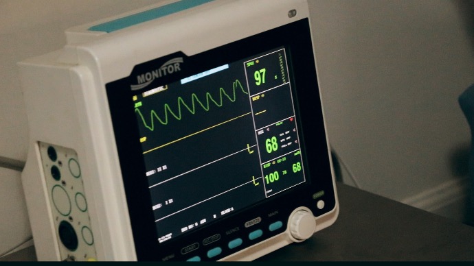
An electrocardiogram (ECG or EKG) is a test that measures the electrical activity of the heart. It is used to detect abnormalities in the heart's rhythm and structure. During an ECG, electrodes are placed on the person's chest, arms, and legs. These electrodes are connected to an ECG machine, which records the electrical activity of the heart.The ECG is a non-invasive test, meaning that it does not involve any needles or incisions. It is generally a painless procedure, although some people may experience some discomfort from the electrodes being placed on the skin. The ECG can provide important information about the heart's function and can help diagnose a variety of conditions, including heart attacks, arrhythmias, and heart disease.The test is typically performed in a doctor's office or hospital and takes about 5-10 minutes.
To prepare for an electrocardiogram (ECG), there are a few things you can do to ensure that the test results are accurate:
- Wear comfortable, loose-fitting clothing.
- Avoid caffeine and other stimulants.
- Avoid lotions and oils on the skin.
- Inform your doctor of any medications you are taking.
- Inform your doctor of any symptoms you may be experiencing.
- Inform your doctor of any medical history.
An echocardiogram (also known as an "echo") is a non-invasive test that uses high-frequency sound waves (ultrasound) to create detailed images of the heart. These images can provide information about the heart's size, shape, and function, as well as the blood flow through the heart and its surrounding blood vessels.
During the test, a special transducer (a small handheld device) is placed on the chest, and sound waves are directed at the heart. The transducer picks up the echoes of the sound waves and sends them to a computer, which creates detailed images of the heart.
There are several different types of echocardiograms, including:
- Transthoracic echocardiogram (TTE): This is the most common type of echo, and is performed with the transducer placed on the chest.
- Transesophageal echocardiogram (TEE): This type of echo is performed with the transducer placed in the esophagus, which allows for a clearer view of the heart.
- Stress echocardiogram: This test is done while the patient is exercising or receiving medication to simulate exercise. This test is done to detect heart issues that are related to exercise or stress.
An echocardiogram (echo) is typically done in a hospital or our facility. The procedure typically takes around 30 to 60 minutes, depending on the type of echocardiogram being performed and the amount of information that needs to be gathered.
A Holter monitor is a type of portable electrocardiogram (ECG) device that is worn by a patient for a period of time, usually 24 to 48 hours, to record the heart's electrical activity. It is used to detect abnormal heart rhythms (arrhythmias) that may not be present during a standard ECG, which is typically only done for a few minutes.
During the test, a small, portable ECG machine is worn by the patient, typically on a belt around the waist or over the shoulder. The device is connected to electrodes that are placed on the patient's chest, and it continuously records the heart's electrical activity over a period of time.
The patient will be instructed to keep a diary of their symptoms and activities throughout the test, to help the doctor determine if there is a correlation between symptoms and the abnormal heart rhythms. After the monitoring period is over, the patient will return the monitor to the doctor and the data will be analyzed. The results of the test will be provided to the patient's physician.
Holter monitoring is a useful diagnostic tool for detecting and evaluating arrhythmias such as atrial fibrillation, ventricular tachycardia, and palpitations. It is also used to evaluate the effectiveness of treatment for these conditions.
Your doctor will check your pacemaker on a regular basis to ensure that it is working properly and that the settings are appropriate for you. Interrogation is the process of checking your pacemaker settings. The pacemaker can be programmed to control the strength and length of the impulse sent to the heart muscle, as well as how fast it will go. If necessary, your doctor may change the pacemaker programming.
An ankle-brachial index (ABI) is a test that measures the blood pressure in your ankle in comparison to the blood pressure in your arm. The test is used to evaluate peripheral artery disease (PAD), which is a condition where the blood vessels in your legs are narrowed or blocked, causing poor blood flow.
The test is performed by measuring the blood pressure in the ankle and arm using a special cuff and a handheld Doppler ultrasound device. The test is usually done on both legs, and the results are compared to each other. The ratio of the ankle blood pressure to the arm blood pressure is calculated, and this ratio is known as the ABI.
A normal ABI is generally considered to be between 0.9 and 1.3. A value below 0.9 indicates PAD, while a value above 1.3 is considered abnormal and may indicate other conditions such as hypertension or arterial calcification.
The ABI test is simple, non-invasive, and painless. It is performed in a clinic or doctor's office, and typically takes about 15 to 20 minutes. It is usually done as part of a regular physical examination, or as a follow-up test after symptoms such as leg pain, cramping, or claudication (painful cramping in the legs and buttocks that occurs during exercise) have been reported.
A carotid ultrasound is a non-invasive test that uses high-frequency sound waves (ultrasound) to create detailed images of the carotid artery in the neck. The carotid artery is one of the main blood vessels that supplies blood to the brain, and the test is used to evaluate the health of the carotid artery and detect the presence of any blockages or narrowing (stenosis) in the artery.
During the test, a special transducer (a small handheld device) is placed on the patient's neck, and sound waves are directed at the carotid artery. The transducer picks up the echoes of the sound waves and sends them to a computer, which creates detailed images of the carotid artery.
The test is typically performed to identify the presence of blockages or stenosis in the carotid artery, which can increase the risk of stroke. The test is also used to evaluate the effects of treatment such as carotid endarterectomy, a surgical procedure to remove the plaque from the carotid artery.
The test is usually done in a clinic or hospital setting and takes around 30 to 60 minutes. The patient will be positioned comfortably on an examination table, and the procedure is usually painless. There is no need for specific preparation but it's important to inform the healthcare provider if you have any medical conditions, allergies or are pregnant.
Peripheral vascular disease (PVD) is a condition that affects the blood vessels in the body outside of the heart and brain. It is also commonly known as peripheral artery disease (PAD). PVD is caused by a buildup of plaque in the blood vessels, which can restrict or block blood flow to the legs, arms, and other areas of the body.
The most common symptoms of PVD include leg pain, cramping, and fatigue during physical activity (claudication). These symptoms are caused by a decrease in blood flow to the legs, and they usually improve with rest. PVD can also cause non-healing wounds, infections or gangrene on the legs or feet.
Diagnosis of PVD is usually made by a physical examination and a test such as an ankle-brachial index (ABI), which compares the blood pressure in the ankle to the blood pressure in the arm. Other tests such as duplex ultrasonography, CT Angiography and MRI Angiography can also be used to visualize the blood vessels and the extent of the blockage.
Medical procedures:
- Angioplasty and stenting: These procedures involve using a catheter with a balloon on the end to open up a blocked blood vessel. A stent, a small mesh tube, is then inserted to keep the vessel open.
- Atherectomy: This procedure involves using a catheter to remove plaque from the inside of a blood vessel.
- Bypass surgery: This procedure involves creating a new pathway for blood to flow around a blocked blood vessel.
It's important to note that the specific treatment plan will depend on the individual patient, the severity of their PVD and other factors such as other underlying medical conditions. Your healthcare provider will work with you to create a treatment plan that is tailored to your needs.
It is also important to regularly follow up with your healthcare provider, to monitor your condition and make any necessary adjustments to your treatment plan. Managing PVD can help prevent serious complications such as amputation, heart attack, or stroke.
Vein disease, also known as venous insufficiency or venous reflux disease, is a condition that affects the veins in the legs and can cause a range of symptoms such as varicose veins, leg swelling, pain, fatigue, and skin changes. The management and treatment of vein disease typically involves a combination of lifestyle changes, compression therapy, and medical procedures.
Lifestyle changes:
- Exercise: Regular exercise, such as walking or cycling, can help improve blood flow in the legs and reduce symptoms.
- Maintain a healthy weight: Excess weight can put extra pressure on the veins in the legs and increase the risk of vein disease.
- Avoid standing or sitting for long periods: Prolonged standing or sitting can cause blood to pool in the legs and increase the risk of vein disease.
- Elevate your legs: Elevating your legs while sitting or lying down can help improve blood flow and reduce swelling.
Compression therapy:
Compression stockings: These are tight-fitting stockings that apply pressure to the legs and help improve blood flow. They are usually worn during the day and can help reduce swelling, pain, and fatigue.
Medical procedures:
Sclerotherapy: This procedure involves injecting a solution into the affected vein, which causes the vein to collapse and disappear.
Endovenous ablation: This procedure uses heat or lasers to seal off the affected vein and redirect blood flow to healthy veins.
Vein ligation and stripping: This procedure involves tying off the affected vein and removing it from the body.
Aortic aneurysm is a bulge in the wall of the aorta, the main blood vessel that carries blood from the heart to the rest of the body. It can be monitored with imaging tests such as ultrasound, CT scan, or MRI. If the aneurysm is large or growing rapidly, it may require repair through surgery, endovascular repair (using a stent-graft), or open surgical repair. The choice of treatment depends on the size, location, and rate of growth of the aneurysm, as well as the patient's overall health.
Preparation for aortic aneurysm surgery includes:
Pre-operative evaluation: Patients undergo medical and imaging tests to determine the best course of action and assess their overall health.
Stop certain medications: Patients may be advised to stop taking certain medications, such as blood thinners, before surgery.
Quit smoking: Patients who smoke are advised to quit as smoking can complicate the healing process.
Discuss the risks and benefits of the surgery: Patients should discuss the risks and benefits of the surgery with their doctor and make an informed decision.
Follow a healthy diet and exercise regimen: Patients are encouraged to maintain a healthy lifestyle, including a balanced diet and regular exercise, to prepare for surgery and the recovery period.
Arrange for post-operative care: Patients should make arrangements for post-operative care, such as transportation and help at home, to ensure a smooth recovery.
High blood pressure and high cholesterol are linked in both directions. When the body is unable to remove excess cholesterol from the bloodstream, it can deposit along artery walls. When arteries stiffen and narrow due to deposits, the heart has to work extra hard to pump blood through them. This causes blood pressure to rise and rise. High blood pressure can damage arteries in its own right over time. It causes tears in the artery walls, allowing excess cholesterol to accumulate. Because the damage they cause over time, high cholesterol and high blood pressure are the two main risk factors for heart disease and stroke.
High blood pressure and cholesterol can be monitored through:
Regular check-ups: Regular check-ups with a healthcare provider are important to monitor blood pressure and cholesterol levels.
Self-monitoring: Self-monitoring of blood pressure using a home blood pressure monitor can be helpful in tracking blood pressure levels between doctor visits.
Blood tests: A blood test called a lipid profile can measure cholesterol levels, including low-density lipoprotein (LDL) cholesterol, high-density lipoprotein (HDL) cholesterol, and triglycerides.
Keeping a record: Keeping a record of blood pressure and cholesterol readings can help track progress and provide important information for doctor visits.
Heart failure is a chronic disease that must be managed for the rest of one's life. However, with treatment, heart failure symptoms and signs can improve, and the heart can sometimes become stronger. Heart failure can sometimes be reversed by treating the underlying cause. Repairing a heart valve or controlling a fast heart rhythm, for example, may reverse heart failure. However, for the majority of people, treating heart failure entails a combination of the appropriate medications and, in some cases, the use of devices that assist the heart in beating and contracting properly.
Heart failure management involves a combination of lifestyle changes, medications, and medical procedures. The specific approach depends on the type and severity of heart failure, as well as other factors such as overall health and age. Some common approaches include:
- Lifestyle changes: Eating a healthy diet, engaging in physical activity, losing weight if necessary, avoiding alcohol and salt, and quitting smoking can help manage heart failure.
- Medications: Prescription medications, such as angiotensin-converting enzyme inhibitors (ACE inhibitors), angiotensin receptor blockers (ARBs), beta blockers, diuretics, and others, can help manage heart failure by improving heart function and reducing fluid buildup.
- Devices: Devices such as implantable cardioverter-defibrillators (ICDs) and heart pumps can be used to improve heart function and prevent heart failure complications.
- Procedures: Procedures such as coronary artery bypass surgery, angioplasty, and heart valve surgery can be used to treat underlying conditions that contribute to heart failure.
- Monitoring: Regular monitoring of symptoms, medications, and heart function is important in managing heart failure.
The procedure of angioplasty is used to open blocked coronary arteries caused by coronary artery disease. It improves blood flow to the heart muscle without the need for open-heart surgery. In an emergency situation, such as a heart attack, angioplasty can be performed. It can also be done as elective surgery if your doctor suspects you have heart disease. Percutaneous coronary intervention is another term for angioplasty (PCI).A stent is a tiny, expandable metal mesh coil. It is put into the newly opened area of the artery to help keep the artery from narrowing or closing again.
Heart catheterization is a diagnostic procedure that involves inserting a thin, flexible tube (catheter) into a blood vessel and guiding it to the heart. The procedure is used to:
-
Assess blood flow: To determine if blood is flowing normally through the heart and blood vessels.
-
Evaluate heart function: To determine how well the heart is functioning and if it is damaged.
-
Diagnose heart conditions: To diagnose heart conditions such as blockages, congenital heart defects, or heart disease.
-
Treat heart conditions: To perform certain treatments such as angioplasty, stent placement, or heart valve repair.
Heart catheterization is typically performed under local anesthesia or conscious sedation and can take anywhere from 30 minutes to a few hours. The procedure has some risks, including bleeding, infection, and damage to blood vessels or heart tissue, but these risks are generally low. Your healthcare provider can provide more information about the risks and benefits of the procedure and what to expect during and after the procedure.
The ankle brachial index (ABI) is a straightforward test that compares blood pressure in the upper and lower limbs. ABI is calculated by dividing blood pressure in an artery of the ankle by blood pressure in an artery of the arm. The ABI is the end result. If this ratio is less than 0.9, a person may have peripheral artery disease (PAD) in the blood vessels in his or her legs.
Write one or two paragraphs describing your product or services.
To be successful your content needs to be useful to your readers.
Start with the customer – find out what they want and give it to them.


PREVENTIVE CARE

Eat a healthy, balance diet.
A healthy and balanced diet is important for preventing cardiovascular diseases. Eat plenty of fruits and vegetables, whole grains, and lean protein. Avoid processed foods, sugary drinks, and excessive amounts of saturated and unhealthy fats.

Be more active
Exercising regularly and maintaining a healthy weight are also important for preventing cardiovascular diseases.

Avoid alcohol and smoking
Smoking is a major risk factor for developing cardiovascular disease. It is also the leading cause of coronary thrombosis in people under the age of 50. If you drink, do not exceed the recommended limit, always avoid binge drinking at all costs, as it increases the risk of a heart attack.

Take prescribed medicine
Whether you have coronary heart disease, high cholesterol, high blood pressure, or a family history of heart disease, it is critical that you take any medication prescribed by your doctor and follow the correct dosage. Do not stop taking your medication without first consulting a doctor.
According to the Centers for Disease Control, heart disease is the leading cause of death for both men and women in the United States.
How Diabetes affects your heart?
Diabetes can have a variety of effects on your heart. For one thing, it increases the risk of developing cardiovascular disease. This is because diabetes can damage blood vessels, causing blockages or narrowing of the arteries. This can then increase the risk of a heart attack or stroke. Diabetes can also increase the risk of developing heart failure. This is because when blood sugar levels are high, the heart has to work harder to pump blood. This can eventually cause the heart muscle to weaken and become inefficient. Finally, diabetes increases the risk of developing abnormal heart rhythms, which can be dangerous and potentially fatal.
Controlling your blood sugar is essential for managing diabetes.
We are in good company
Our providers perform procedures at any of these locations.


